

HEALTH INFORMATION
Maintaining good health care for your dog is one of the most important obligations all owners face. As a breeder our puppies will receive their initial vaccinations which include Vangaurd 5 way DHLPP and 4-5 dewormings of Nemex-2. Cold Mountain Siberians takes pride in the quality of our dogs and provides a full health guarantee at the time of sale however there are many diseases and illnesses that your dog may come into contact with in the future. Take the time to familiarize yourself with illnesses commonly found in dogs.
Heartworm
Whether or not you choose to use conventional heartworm preventatives, there is always the possibility that you may one day find yourself with a heartworm-positive dog, and be faced with the decision of how to treat it.
There are a number of reasons why a dog becomes heartworm-positive. One of the most common is adopting a dog from a rescue organization. Especially in the South where heartworm is ubiquitous, most dogs that are not given heartworm preventative regularly will be test positive for heartworm. There is also a small chance that your dog could become infected with heartworms if you choose to extend the time between doses of the heartworm preventative you give your dog beyond what is recommended on the label, if you give less than the recommended dose, or if your dog should vomit up the pill without your knowledge. It is important to understand that heartworm infections are not detectable until about six months after a dog has been bitten by a heartworm-infected mosquito. Blood tests generally will not detect heartworms in a dog until the larvae have matured into adult worms, which takes about six months following initial infection. Symptoms, such as coughing, lethargy and difficulty breathing, will not show up until the infection is advanced.
HOW DID HEARTWORM IS CONTRACTED- Heartworm disease is caused by an infestation of a parasite, Dirofilaria immitis, commonly called heartworm, with an elaborate life cycle. It starts in an infected animal; more than 30 species, including dogs and wild animals such as coyotes, foxes, and ferrets act as "reservoir" species. Adult worms, residing in the host animal's heart, lungs, and associated blood vessels, mate and the females release their young (called microfilariae). These circulate in the host animal's blood for up to two years. They develop into their next stage of life, L1 (for first larval stage) only if ingested by a mosquito during a mosquito's blood meal. It takes the L1 larvae 8 to 28 days, depending on environmental temperatures, to develop into their third stage (L3), when they migrate from the mosquito's stomach to its mouth. The L3 larvae enter their next host through the mosquito's next bite. As many as 10 to 12 L3 larvae can be transmitted to a dog in a single mosquito bite. The L3 larvae molt and migrate through the dog's tissues in search of major veins, which they infiltrate and use as a path to reach the heart. It takes them about 90 to 100 days to develop into L5, the form that breaches the circulatory system.
STAGES- Heartworm infection is divided into four or five stages (depending on the model used), based on the severity of the infestation and the age and health of the dog.
Stage 1 (mild)- consists of young, healthy dogs with no symptoms and minimal changes evident on X-rays.
State 2 (moderate)- infection will show heartworm disease that is evident on X-rays, but symptoms are minimal, mostly coughing.
Stage 3 - a severe infection, with weight loss, coughing, difficulty breathing, more damage visible on X-rays, along with liver and/or kidney damage.
Stage 4 and 5- are considered critical, with the dog often collapsing in shock. These dogs will not survive ordinary heartworm treatment, and must have the worms surgically removed if they are to have any hope of survival.
SYMPTOMS OF INFECTION:
1. Mild disease: Cough
2. Moderate disease: Cough, exercise intolerance, abnormal lung sounds
3. Severe disease: Cough, exercise intolerance, difficulty breathing, abnormal lung sounds, enlargement of the liver, temporary loss of consciousness due to poor blood flow to the brain, fluid accumulation in the abdominal cavity, abnormal heart sounds, death.
*The 2005 Guidelines for the treatment of heartworm recommend starting dogs on heartworm preventative immediately.
If you feel your dog may be heartworm-positive please contact your local veterinarian immediately.


Giardia (Giardiasis)
LIFE CYCLE- Giardia occur in two forms: a motile feeding stage that lives in the intestine, and a non-motile cyst stage that passes in the feces. The giardia trophozoite, which is the active stage of the organism - inhabits the small intestine of the dog. The trophozoite stage is tear-drop shaped, binucleated, and has four pairs of flagella. It attaches to the cells of the intestine with its adhesive disc and rapidly divides to produce a whole population of trophozoites. As they detach they may be swept down the intestine. If intestinal flow is fast then they may appear in the feces. However, if they have time, encystment occurs as the parasite travels to the large intestine. The cyst is fairly resistant, and can survive for several months outside of a host's body as long as sufficient moisture is provided. Mature cysts are usually found in the feces of infected animals. Other animals become infected by ingesting the cysts that passed from the body in feces. These ingested cysts then break open inside the small intestine to release the motile feeding stage. Giardia increase their numbers by each organism dividing in half.
HOW GIARDIA IS CONTRACTED- Giardia infections are transmitted via ingestion of trophozoites or cysts in contaminated water or food. Transmission also occurs by direct contact, especially with asymptomatic carriers. More recently, giardiasis has also been recognized as being able to be sexually transmitted. Giardia is so prevalent throughout North America because it is highly contagious. The ingestion of as few as one or more giardia cysts may cause the disease.
SYMPTOMS- Clinical signs range from none in asymptomatic carriers, to mild recurring diarrhea consisting of soft, light-colored stools, to acute explosive diarrhea in severe cases. Other signs associated with giardiasis are weight loss, listlessness, fatigue, mucus in the stool, and anorexia. These signs are also associated with other diseases of the intestinal tract, and are not specific to giardiasis. These signs, together with the beginning of cyst shedding, begin about one week post-infection. There may be additional signs of large intestinal irritation, such as straining and even small amounts of blood in the feces. Usually the blood picture of affected animals is normal, though occasionally there is a slight increase in the number of white blood cells and mild anemia. Without treatment, the condition may continue, either chronically or intermittently, for weeks or months.
Thorough cleaning of all kennel areas where infected dogs have access is essential. Once organic debris has been removed, thorough disinfection will help to further reduce the level of environmental contamination and reduce the risk of dogs becoming re-infected after the completion of treatment.
*If you feel your dog has contracted Giardia please contact your local veterinarian immediately.

Giardiasis is an intestinal infection caused by a parasitic single celled organism called Giardia lamblia. These organisms are found in the intestines of many animals, including dogs and humans. This microscopic parasite clings to the surface of the intestine, or floats free in the mucous lining the intestine.
Coccidiosis
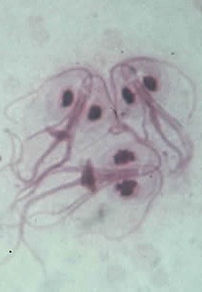
Coccidiosis is an intestinal disease that affects several different animal species including canines and humans. Coccidia is one of the most prevalent protozoal infections in North American animals, second only to Guardia.
Eimeria and Isospora are the two genera that are often referred to as "coccidia." These two genera contain a large number of species that infect a variety of animals throughout the world. The diseases caused by these microscopic protozoal parasites are referred to collectively as Coccidiosis, and they vary tremendously in virulence. Some species cause diseases that result in mild symptoms that might go unnoticed (I.e., mild diarrhea) and eventually disappear, while other species cause highly virulent infections that can be rapidly fatal. The causative agent is a protozoan that has the ability to multiply rapidly. The major damage is due to the rapid multiplication of the parasite in the intestinal wall, and the subsequent rupture of the cells of the intestinal lining. Several stages of multiplication occur before the final stage, the oocyst, is passed in the feces. Oocysts are extremely resistant to environmental stress and are difficult to completely remove from the environment. Oocysts are frequent contaminants of feed and water and when the sporulated Oocysts are ingested by other animals they start the life cycle over in the new host.
LIFE CYCLE- The life cycles of both genera of coccidia are similar. A host is infected when it ingests Oocysts that have been passed in the feces of another host. The oocyst encysts in the host's small intestine, and the sporozoites contained within the oocyst are liberated. The sporozoites penetrate the cells of the host's small intestine and reproduce asexually. Each generation of asexual reproduction produces multiple merozoites; the merozoites are liberated from the cell and infect new cells. It is this stage of the infection that can result in destruction of massive numbers of cells in the host's small intestine and, ultimately, lead to the host's death. Death may follow the acute disease either directly or from secondary diseases s uch as pneumonia. Animals that survive for 10 to 14 days may recover, however, permanent damage may occur. The susceptibility of animals to this disease varies. The ingestion of Oocysts may not produce the disease; some animals constantly carry them without being affected. Recovered animals develop immunity and seem to be partially resistant to reinfection.
SYMPTOMS- Clinical signs of Coccidiosis usually are present or shortly following stress such as weather changes; weaning; overcrowding; long automobile or plane rides; relocation to a new home and new owners; and/or unsanitary conditions. Symptoms or signs of Coccidiosis will depend on the state of the disease at the time of observation. In general, Coccidiosis affects the intestinal tract and symptoms are associated with it. In mild cases, only a watery diarrhea may be present, and if blood is present in the feces, it is only in small amounts. Severely affected animals may have a thin, watery feces with considerable amounts of intestinal mucosa and blood. Straining usually is evident, rapid dehydration, weight loss and anorexia (off food) also may be clinically visible. "Nervous Coccidiosis" is a nervous system condition associated with coccidial infection. Signs are consistent with central nervous system involvement, and include muscle tremors, convulsions and other central nervous system symptoms. A consistent sign in "nervous cocci" dogs is that stimulation of any type seems to trigger the symptoms.
*If you feel your dog may have contracted Coccidiosis please contact your local veterinarian immediately.

Distemper
Canine distemper is a contagious and serious viral illness with no known cure. The disease affects dogs, and certain species of wildlife, such as raccoons, wolves, foxes, and skunks. The common house pet, the ferret, is also a carrier of this virus. Canine distemper belongs to the Morbillivirus class of viruses, and is a relative of the measles virus, which affects humans, the Rinderpest virus that affects cattle, and the Phocine virus that causes seal distemper. All are members of the Paramyxoviridae family. Young, unvaccinated puppies and non-immunized older dogs tend to be more susceptible to the disease.
HOW DISTEMPER IS CONTRACTED- The infected dog typically infects other dogs via coughing infected respiratory secretions though the virus is shed in most other body secretions including urine. The distemper virus consists of a single strand of RNA, encased in a protein coat which is again encased in a fatty envelope. This sounds esoteric but the fatty envelope makes all the difference in the world. The fatty envelope is easily disrupted in the environment which makes it impossible for infectious virus to persist in the environment. Because an intact fatty envelope is required for infection, virus transmission must involve dog to dog contact or at least contact with extremely fresh (less than 30 minutes old at 60 degrees and up to 3 hours old at room temperature) infected body secretions. As with other viruses, living virus happily freezes and can survive for years if kept frozen and protected from light. Routine disinfection and cleaning readily kills the distemper virus in the kennel setting.
LIFE CYLCE- The virus enters the new host via the nose or mouth and promptly begins to replicate. Virus is engulfed by cells of the immune system called “macrophages.” The idea is that the virus will be engulfed, walled off within the cell and then destroyed by enzymes. Unfortunately for the new host, this process does not damage the virus as intended; instead, the virus is able to use the macrophage as a means of transportation through the host’s body. Within 24 hours, the virus has traveled to the lymph nodes of the lung. By the 6th day, the virus has migrated to the spleen, stomach, small intestine, and liver. Fever is developing at this point.
By day 8 or 9 an important crux is reached in the timetable of infection. The host is mounting an immune response during this time and the outcome depends on how fast and how well this is accomplished. A strong immune response begins to clear the virus at this point and has eliminated all traces of virus with no symptoms of illness by Day 14. A weak immune response allows the virus to reach the ”epithelial cells,” the cells which line every interface the body has with the outside world. The tender epithelial cells lining the chambers of the brain are infected as well. The host begins to get sick as the virus spreads but as the host's immune response grows symptoms wane. This phenomenon accounts for the wide variability in symptoms; some dogs get only a few mild symptoms while others get a full lethal combination.
After clearing from most internal organs, the virus is able to “hideout” for long periods of time in the nervous system and skin. Because of this phenomenon, callusing of skin or, much worse, seizures may occur long after the infection was thought to be cleared.
Most victims in the U.S. are puppies. (The colostrum suckled in the first day or so of life will provide them with a solid reflection of their mother’s immunity. This will have waned by age 16 weeks leaving the puppy vulnerable if vaccines have not been administered for further protection. In our society most mother dogs will have received some form of vaccination and thus be able to pass on at least some immunity and will have some ability to protect herself. In societies where vaccination is not common, distemper attacks all age dogs.)
SYMPTOMS- begin with:
• Gooey eye and nose discharge
• Fever (which often comes and goes unnoticed)
• Poor appetite
• Coughing and the development of pneumonia
The virus is attacking interfaces of the body with the environment (the mucous membranes) and starts with the respiratory tract, hence the pneumonia, but it does not stop there. The virus moves on to produce:
• Vomiting and diarrhea
• Callusing of the nose and foot pads (hence one of the old names for distemper - hard pad disease)
After completing what is called the “mucosal phase” of infection where environmental interfaces are attacked (as described by the above GI and respiratory disease), the virus proceeds to the central nervous system for its “neurologic phase” leading to:
Seizures (classically starting with snapping or tremoring of the jaws that progress to convulsions of the whole body. (This distemper classic sign is called a chewing gum fit.)
• Seizures are not the only distemper sign by any means. Tremors , imbalance, and limb weakness all may occur. Signs may progress to death or may become non-progressive and permanent. Recovery is also possible.
This means that the dog appears to recover only to break with neurologic disease 1 to 3 weeks later. Younger puppies or individuals with weak immunity often die during the mucosal phase while stronger individuals may have relatively mild mucosal signs and not appear ill until the neurologic phase strikes.
The canine distemper virus is closely related to the human measles virus and, in fact, in older times, puppies were immunized for distemper with vaccine against measles.
*If you feel your dog may have been infected with Distemper please contact your local veterinarian immediately.


